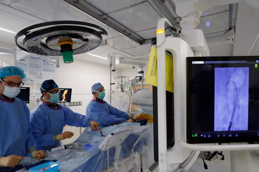
Photo above: (Left to right) Vascular Surgeon Douglas Jones, MD, and PG5 resident Zachary Fang, MD join Andres Schanzer, MD, FACS in performing a complex FEVAR procedure.
Endovascular repairs are done in a hybrid cardiovascular operating room with advanced imaging equipment, a fluoroscopy unit and traditional operating suite. At UMass Memorial Medical Center, the hybrid OR is also equipped with Fiber Optic RealShape (FORS) technology. Surgeons there were the first in the nation to use FORS for a complex endovascular aortic aneurysm repair (EVAR). The technology gives operators better imaging and more flexibility in maneuvering endovascular guidewires and catheters for EVAR, while also reducing dependence on fluoroscopy.
Most vascular surgeons rely on two-dimensional, grayscale fluoroscopy imaging to visualize endovascular devices. For the patient, the radiation exposure can be significant, although the need for the procedure outweighs that risk. For surgeons and other members of the clinical team who conduct the procedures repeatedly, the radiation exposure has the potential to become more consequential as it accumulates during training programs and the procedures themselves.
A new technology developed by Philips dramatically improves visibility while also reducing dependence on fluoroscopy. In 2021, UMass Memorial Medical Center vascular surgeons became the first in the nation to use FORS, a technology that generates real-time images of endovascular devices by refracting light through optical fibers. Only two other hospitals in the U.S. and three outside the country are currently using it.
Reducing radiation exposure
“Radiation continues to be an essential tool for us, but we all know it can pose a threat to the health of a patient and to everyone on the treatment team,” said Andres Schanzer, MD, FACS, Chief, Division of Vascular and Endovascular Surgery, Director, Center for Complex Aortic Disease at UMass Memorial Medical Center, Director, UMass Memorial Health Heart and Vascular Center, and Professor, UMass Chan Medical School.
Instead of the ionizing radiation of fluoroscopy, FORS technology uses light refracted through optical fibers to provide 3D tracking of endovascular guidewires, catheters and devices. FORS technology has the potential to reduce a patient’s radiation exposure by as much as 75% during endovascular complex aortic aneurysm repair.
“FORS technology gives us the opportunity to protect our patients and colleagues from radiation exposure, while providing safe, effective, minimally invasive repairs with even greater accuracy,” Schanzer said.
Imaging advantages
In addition to reducing the need for fluoroscopy, FORS technology enhances the visibility as the operator moves devices through the arteries. The real-time images produced are three dimensional, so the EVAR team can view progress and movement from any angle. The technology also allows for zooming in and having multiple projections on-screen at once.
“This enables me to always keep the full extent of the guidewire in my field of vision,” Schanzer said. “Then I can advance the FORS catheter over the wire to get it in position. The FORS system shows which portion of the guidewire or catheter is in front of the other, making all endovascular interventions easier and safer.”
The FORS images are displayed on top of, and in alignment with, anatomical patient images created through digital subtraction angiography (DSA) or computed tomography (CT), both of which are black and white. With DSA or CT images alone, it can be difficult to determine the exact placement of the catheter, but with FORS technology, it can be seen in color in the foreground.
“The system is very intuitive and truly represents a new era in device guidance,” Schanzer said, adding that having a color image has an additional benefit. “Until I started using FORS, I hadn’t realized how much operator fatigue there is when you’re only looking at different shades of gray to visualize everything. It’s become very apparent to me that the bright yellow and blue on top of the gray background really decreases that strain, especially when you’re looking at previous stent grafts and wires.”
View more stories of healing, advancing medicine and innovating from UMass Memorial Health.